Claudia's Projects
Gaze-Contingent Calibration
Microsaccade Correction During Sustained Fixation
1. Gaze Contingent Calibration
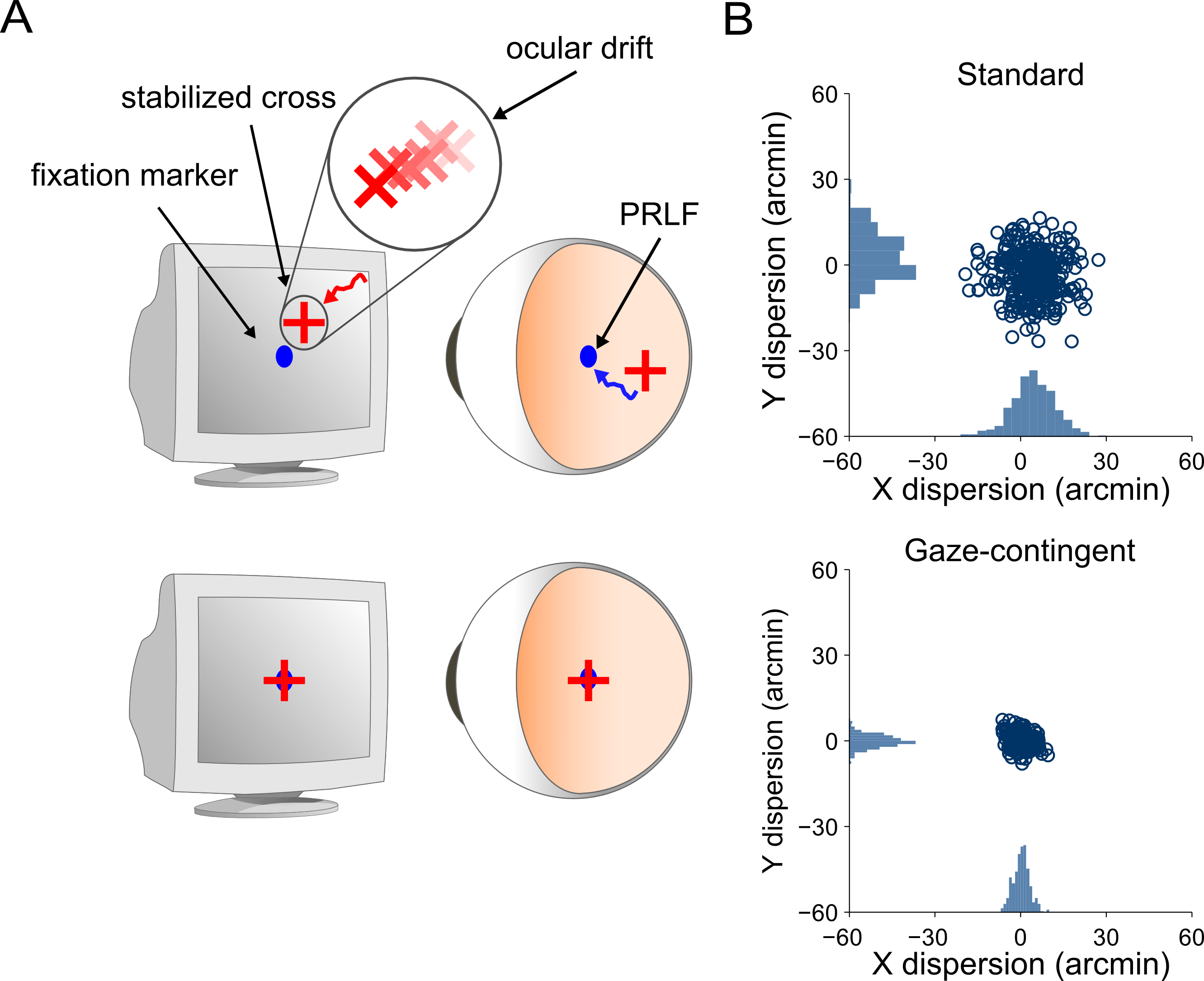
This database was collected to quantify the effect of a gaze-contingent calibration. The experiment was developed to mimic the calibration procedure used by EyeRIS. The figure reports data from an observer.

Gaze-contingent calibration procedure.
a: Schematic representation of the gaze-contingent calibration procedure. A fixation marker (blue dot) and the position of gaze estimated by the eye tracker (red cross) are displayed on the CRT monitor. Subjects adjusted the offsets of the estimated gaze position by means of a joypad while maintaining fixation on a marker.
b: Dispersion of gaze in the standard one-step calibration procedure used in oculomotor research (top) and in our method (bottom).
Subjects
Two observers with normal, non-corrected vision participated in the original study. All observers had had previous experience with psychophysical experiments requiring sustained fixation and they were all naïve about the purposes of the experiment.
Apparatus and Stimuli
The experiment was conducted in complete darkness. Stimuli were displayed on a fast phosphor monitor (Iyamaya HM204DT) at a resolution of 800x600 pixels and vertical refresh rate of 200 Hz.
Procedure
Every session started with preliminary setup operations that lasted a few minutes and allowed the subject to adapt to the low level of light in the room. Preliminary operations included: (1) positioning the subject optimally and comfortably in the apparatus; (2) tuning the eyetracker; and (3) performing a calibration procedure to convert the voltages given by the eyetracker into degrees of visual angle. These operations were repeated before each block of trial.
The experiment consisted in alternating two conditions. In the first condition (automatic calibration) observers sequentially fixated at the center of crosses placed on a grid of nine equispaced points, as standard calibration procedure. Observers were asked to press a button of the joypad when they were fixating at the center of the cross.
Data And Analysis
The data and analysis can be found at: \\opus.cvs.rochester.edu\aplab\ClaudiaProjects\MicrosaccadeCorrection
2. Microsaccade Correction During Sustained Fixation
This database was collected because the previous sustained fixation database did not provide precise information of absolute gaze position. In the experiment, trials were collected in three different conditions (dot, dim, dark) were collected consecutively. Twenty or so trials were collected in each condition (???). Unlike the data from the previous sustained fixation database, these data were collected in complete darkness (except for the stimulus on the monitor). The dim condition was inserted to avoid dark adaptation. The data described in the manuscript in process (LINK) refer exclusively to the dot condition.

Schematic representation of the experimental design.
Sequence of trials. Observers were asked to maintain fixation on a dot (in the dot condition) or at the center of the display (in the dim and dark conditions). A calibration procedure was conducted at the beginning of each trial. In the calibration, while fixating on a central marker, subjects corrected the horizontal and vertical offsets of a gaze contingent point displayed at the preferred retinal locus (PRL).
Subjects
Six observers with normal, non-corrected vision participated in the study. All observers had had previous experience with psychophysical experiments requiring sustained fixation and, with the exception of one of the authors, they were all naïve about the purposes of the experiment.
Subject |
Age |
Sex |
S1 |
29 |
F |
S2 |
20 |
F |
S3 |
25 |
M |
S4 |
41 |
M |
S5 |
31 |
M |
S6 |
30 |
F |
Apparatus and Stimuli
The experiment was conducted in complete darkness. Stimuli were displayed on a fast phosphor monitor (Iyamaya HM204DT) at a resolution of 1024x768 pixels and vertical refresh rate of 150 Hz. Subjects were kept at a fixed distance of 123 cm from the monitor. A dental imprint bite bar and a head rest prevented movements of the head. The movements of the right eye were measured by means of a Generation 6 Dual Purkinje Image (DPI) eyetracker (Fourward Technologies). The internal noise of this system is about 20" (Crane and Steele, 1985), enabling a spatial resolution of approximately 1'(Stevenson and Roorda, 2005). Vertical and horizontal eye positions were sampled at 1 KHz and recorded for subsequent analysis. Since we only measured movements of the right eye and used a gaze-contingent calibration procedure to improve localization of the line of sight, stimuli were observed monocularly, with the left eye patched.
For a more detailed description of the beneficial effect of a gaze-contingent calibration procedure see Gaze-contingent Calibration Procedure above.
Procedure
Observers were asked to maintain accurate fixation for 10 s either in the presence (dot condition) or in the absence (dim and dark condition) of a high contrast 4' fixation marker displayed at the center of the monitor. In the dot and dark condition the background of the monitor was uniformly black, whereas it was uniformly gray in the dim condition (monitor contrast: 127, 127, 127). Data were collected in separate experimental sessions, each of approximately one hour duration. Every session started with preliminary setup operations that lasted a few minutes and allowed the subject to adapt to the low level of light in the room. Preliminary operations included: (1) positioning the subject optimally and comfortably in the apparatus; (2) tuning the eyetracker; and (3) performing a calibration procedure to convert the voltages given by the eyetracker into degrees of visual angle. These operations were repeated before each block of trial. Subjects were then presented with four blocks of about 20 experimental trials. Breaks between consecutive blocks ensured that subjects were never constrained in the apparatus for more than 10-15 minutes consecutively.
Data
Data were collected between 2011 and 2012. The original .eis files are in the observers' folders in: \\opus.cvs.rochester.edu\aplab\ClaudiaProjects\FixationPrecision.
No gamma correction was applied to the monitor. Monitor settings were:
• contrast: 70
• brightness: 60
The original .eis files contain the following variables:
• observer's name: "Subject_Name"
• trial duration: "TimeStimulus"
• contrast of the background: "BackgroundGray"
• contrast of the fixation marker: "StimulusGray"
• moment in which the observer refined the calibration before each trial: "ButtonPress"
• experimental condition: "Trial_Type"
The "Trial_Type" coding was:
• 0 = dot condition
• 1 = dim condition
• 2 = dark condition
ValidTrials for each observers can be found in the following folder: \\opus.cvs.rochester.edu\aplab\ClaudiaProjects\FixationPrecision. In the folder the ValidTrials for each observer are categorized according to the experimental condition (e.g. ValidTrialsDotS1: ValidTrials in the dot condition for observer S1).
- • TO DO:
original data .eis and brief documentation on how they have been converted in validtrials
add link to dataset (validtrials) with documentation of how data are organized
add offset
add beginning of the trial
Data Analysis
The .mat (work in progress) codes to generate the figures can be found at: \\opus.cvs.rochester.edu\aplab\ClaudiaProjects\MicrosaccadeCorrection
Notes
8/8/2014 Report: (broken link)
